
Figure 3.
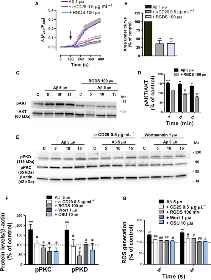
Aβ‐oligomers mobilize intracellular Ca2+ via β1‐integrin and activate β1‐integrin/PI3K signaling in astrocytes. (A, B) Ca2+ responses to Aβ 1 μM in astrocytes freshly isolated from hippocampal sections from P12 rats. Inhibitory effects of RGDS peptide (100 μM) and antibody αCD29 (0.5 μg ml−1) on astrocyte [Ca2+]i following Aβ oligomer treatment (100%). (C, E) Levels of phosphorylated AKT, PKC, and PKD and total AKT and β‐actin were measured by Western blot analysis of astrocyte protein extracts untreated or treated with Aβ (5 μM, 5, 10 and 15 min), RGDS peptide (100 μM), αCD29 (0.5 μg ml−1), wortmannin (1 μM), and OSU‐03012 (10 μM). (D, F) Histograms represent the signal intensity of phosphorylated protein bands after normalization with intensities of total AKT or β‐actin proteins and expressed as a percentage of untreated cells or inhibitor‐treated cells as control values (100%). (G) Effect of β1‐integrin, PI3K, and PDK1 inhibitors on Aβ‐induced ROS levels on cultured astrocytes. *P < 0.05, **P < 0.01, ***P < 0.001 compared to nontreated cells; # P < 0.05, ## P < 0.01 compared to Aβ‐treated cells.