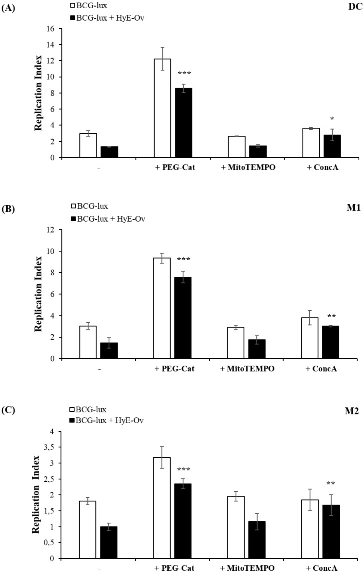
Fig 4. HyE-Ov promotes intracellular mycobacterial killing by a pH and ROS-dependent mechanism.

DC (A), M1 (B) and M2 (C) cells (5x10⁵/well) were infected with BCG-lux for 3 hours (MOI 5) and stimulated or not with HyE-Ov (3 mg/ml of equivalent plant material) in the presence or absence of 100 U/ml PEG-Catalase, 10 μM MitoTEMPO or 10 nM Concanamycin A. Mycobacterial growth was evaluated by luminometric analysis after 3 days (t3) from stimulation (t0). Data are shown as means ± SD of the ratio t3/t0 of RLU values from triplicate cultures and are representative of at least 2 independent experiments performed on cells from different donors. *p<0.05, **p<0.01 and ***p<0.0001 in comparison with HyE-Ov stimulated infected cells.
