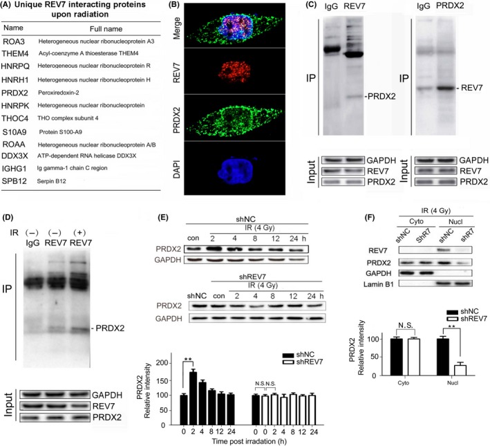
Figure 5.

REV7 interacts with PRDX2 in the nucleus of esophageal squamous cell carcinoma cells. A, List of unique proteins that increased in percentage based on proteomic analysis at 2 h after 4 Gy X‐ray irradiation. B, Immunofluorescent staining of REV7 and PRDX2 in Eca‐109 cells (coexistence of REV7 and PRDX2 are indicated by the arrows). C, Confirmation of PRDX2/REV7 protein co‐immunoprecipitation by beads containing REV7/PRDX2 antibody using whole cell lysate from Eca‐109 cells, as revealed by western blot analysis. D, Immunoprecipitation assay by beads containing REV7 antibody in whole cell lysate from Eca‐109 cell with or without irradiation (2 h after 4 Gy X‐ray), as revealed by western blot analysis. E, Western blotting analysis of PRDX2 expression of shNC/shREV7 group postirradiation. Relative intensity of PRDX2 normalized to GAPDH is presented as the mean ± SEM of 3 independent experiments. **P < .01, N.S. nonsignificant. F, Cytosolic and nuclear fractions isolated from shNC/shREV7 Eca109 cells were assayed by western blotting for PRDX2. Relative intensity of PRDX2 normalized to GAPDH is presented as the mean ± SEM of 3 independent experiments. **P < .01, N.S. nonsignificant
