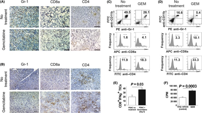
Figure 4.

Effect of gemcitabine (GEM) on tumor‐infiltrating inflammatory cells (TICs) in pancreatic ductal adenocarcinoma (PDAC) models. Intraperitoneal‐dissemination PDAC mice and liver‐metastasis PDAC mice received a single dose of GEM on days 26 and 36, respectively. Two days later, tumor tissues were obtained and used for immunohistochemical analysis and isolation of TICs. A, B, Tumors immunohistochemically analyzed for Gr‐1+, CD8a+ and CD4+ inflammatory cells. Magnification: ×100. Scale bars: 100 μm. A, Intraperitoneal‐dissemination PDAC mice. B, Liver‐metastasis PDAC mice. C, D, FACS analysis of TICs isolated from tumors from intraperitoneal‐dissemination and liver‐metastasis PDAC mice treated with or without GEM. C, TICs isolated from intraperitoneal‐dissemination PDAC mice. D, TICs isolated from liver‐metastasis PDAC mice. One representative scatterplot or histogram in each group is presented. E, TICs were isolated from GEM‐treated (n = 4) or untreated (n = 4) liver‐metastasis PDAC mice, CD8+ cells were selectively sorted from the TICs and stimulated for 8 d with interleukin‐2. Then, IFN‐γ secretion assay was performed. F, The cytotoxicity of CD8+ cells isolated from splenocytes of liver‐metastasis PDAC mice treated (n = 3) or not treated (n = 3) with GEM was assessed by quantifying the amount of [51Cr] released from PAN02 target cells; the Student's t‐test was performed to obtain the P‐value
