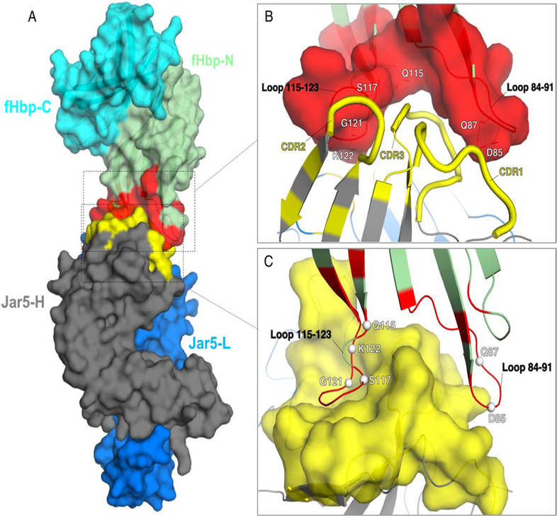
Figure 1. Overall fold and epitope-paratope interface of the complex fHbp:Fab JAR5.
A) The structure of the complex is depicted in a surface representation, with fHbp on top and colored in cyan and green for the C- and N-terminal domains, respectively, while the epitope for JAR5 is colored in red. Heavy and light chains of Fab JAR5 are colored in dark grey and blue, while the paratope is colored in yellow. B) and C) Closer views of the epitope-paratope interface, with the surfaces of the epitope and of the paratope shown in red and yellow. Epitope residues that directly contact JAR5 are labelled white, and shown as lines in B), and as spheres in C).

