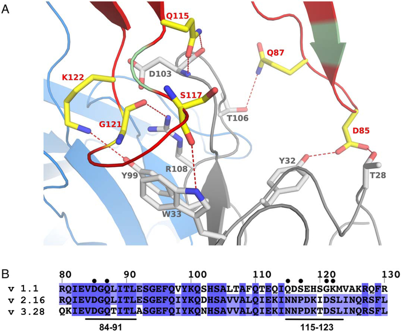
Figure 2. Interactions at the interface between fHbp and JAR5.
(A) Cartoon of fHbp-N, JAR5-L, and JAR5-H, are shown in green, blue, and grey, respectively. Red-color on fHbp-N show all regions interacting with JAR5, while yellow sticks show fHbp residues (labeled in red) involved in direct polar bonds with residues of JAR5-H (shown with grey sticks and grey labels). Direct bonds are indicated with red dashed lines. (B) Sequence alignment of fHbp variants 1.1, 2.16 and 3.28, with the JAR5 epitope annotated with black bars on bottom and labelled. Residues of fHbp v1.1 involved in direct interactions with JAR5 are marked with black circles on top.

