
Figure 5 -. Acid inhibits mTORC1 and the clock in an RAG-independent manner not fully rescuable by TSC2 loss.
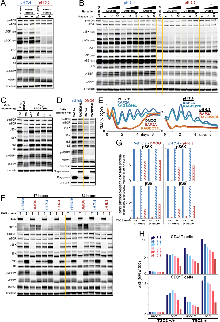
A./B. mTORC1 signaling in U2OS cells unstarved or starved of serum for 50 minutes in pH 7.4 or 6.3 media and then rescued or not for 10 minutes with serum (A), or likewise starved of leucine, arginine, or lysine and rescued with approximately twice the amino acid sensor Km (Wolfson and Sabatini, 2017) or full DMEM level (B). C. mTORC1 signaling in RAP2A- or RAGBQ99L-expressing U2OS Arntl::dLUC cells after 1 h of deprivation of amino acids (AA) or leucine (L) or incubation in replete media. D. Immunoblots of lysate from cell lines in panel C, 23 h after synchronization and treatment with vehicle or 500 uM DMOG in low buffer media. E. Arntl::dLUC luminescence in parallel to D. Mean of 3 BR. F. Immunoblots for HIFIα, mTORCI signaling, and BMAL1 in Arntl::dLUC TSC2 CRISPR knockout (--) or parental Arntl::dLUC U2OS cells (++) following treatment with vehicle or 500 uM DMOG in low buffer media or with pH 7.4 or 6.3 media for 17 and 24 h. RE of 2. G. Quantification of F. Ratio of the intensity of pS6K or pS6 to total S6K or S6, respectively. Each control-treatment pair normalized to respective control (vehicle, pH 7.4). H. Mean fluorescence intensity (MFI) of pS6 staining of wildtype (+/+) and TSC2 knockout (−/−) CD4+ and CD8+ T cells assessed by flow cytometry after TCR stimulation for 1 h in media of the indicated pH. RE of 5 each with 1–4 BR. RE=representative experiment, BR=biological replicates. See also Figure S5.