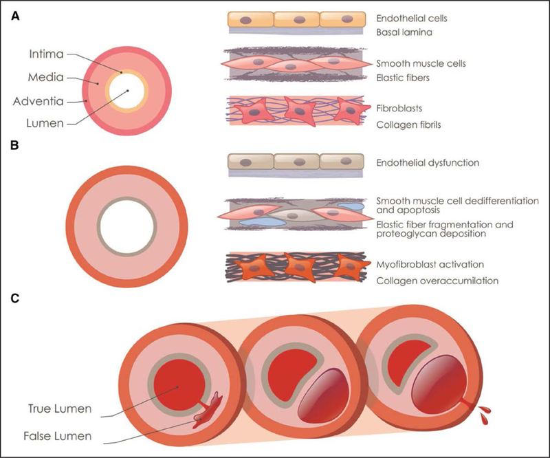
Figure 1.

Pathology and progression of thoracic aortic aneurysms and dissections. A, Schematic illustration of the cellular and ECM (extracellular matrix) components in the 3 layers of the thoracic aorta. B, Cellular and ECM changes associated with aneurysm progression are illustrated, including endothelial dysfunction, elastin fiber fragmentation and loss, increased proteoglycan accumulation (blue), and smooth muscle cell loss (gray cell). C, Illustration of an acute aortic dissection because of a tear in intimal layer, progressing through the medial layer to form a false lumen and rupturing from the false lumen through the adventitial layer.
