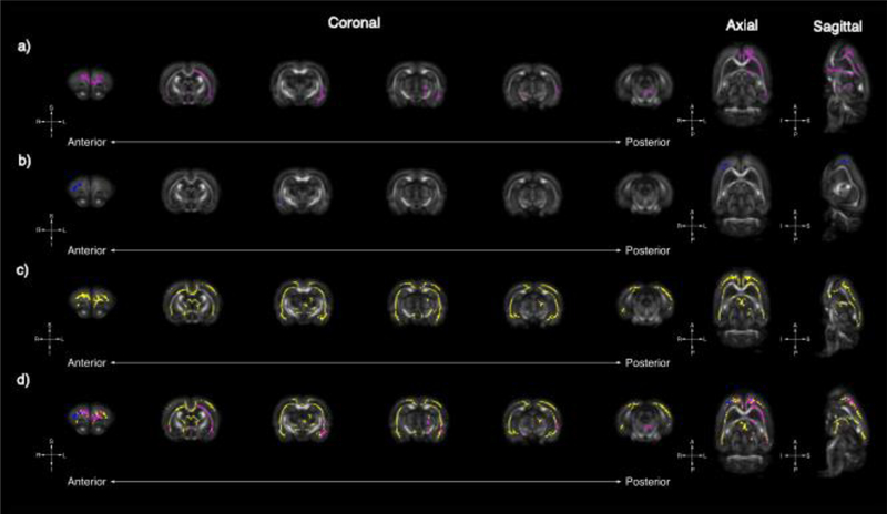
Figure 1.
Tract-based spatial statistics uncovers global changes in RD in Fmr1, Nrxn1, and Pten models of ASD. Whole-brain voxel-wise tract-based spatial statistics reveal significant areas of decreased RD in Fmr1 (A, pink), Nrxn1 (B, blue), and Pten (C, yellow) models of ASD. Six representative coronal sections (left [anterior] to right [posterior]) reveal significant overlapping regions of decreased in RD for all three models in the frontal lobe (D); however, unique gene-specific areas of RD change are noted caudally. Uniquely observed in the Fmr1 model are decreases in RD in the left external capsule whereas unique changes in the Pten model are observed in the frontal lobe, genu of the corpus callosum, and fimbria.

