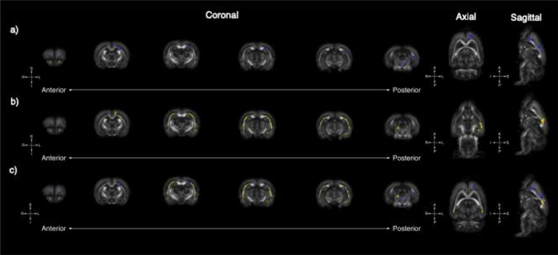
Figure 2.
Tract-based spatial statistics reveals global changes in FA in Nrxn1 and Pten models of ASD: Whole-brain voxel-wise tract-based spatial statistics reveal significant areas of increased FA in Nrxn1 (A, blue) and Pten (B, yellow) models of ASD. Six representative coronal sections (left [anterior] to right [posterior]) reveals unique FA changes in the Nrxn1 model, which are observable medially in anterior regions encompassing the frontal lobe as well as the medial structures of the brain stem. In contrast, increases in FA in the Pten model are most concentrated bilaterally along medial margins of the external capsule. There are also overlapping small areas of increased FA medially in the external capsule and frontal lobe in both the Nrxn1 and Pten models (C).

