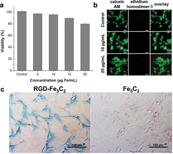
Figure 3.
a) MTT assays with Fe5C2 nanoparticles on U87MG cells. The cells retained over 80% viability in the tested concentration range (0 – 25 μg Fe/mL). b) Live/dead assays with Fe5C2 nanoparticles on U87MG cells. No red fluorescence (ethidium homodimer-1) was observed in the tested concentration range (0 – 25 μg Fe/mL). c) Cellular uptake studies with U87MG cells. The cells were incubated with either RGD-Fe5C2 or Fe5C2 nanoparticles (5 μg Fe/mL) for 1h, and then subjected to Prussian blue staining. Positive staining was found with almost all the cells when RGD-Fe5C2 nanoparticles were used. On the contrary, Fe5C2 nanoparticles showed little cellular uptake.

