Figure 4.
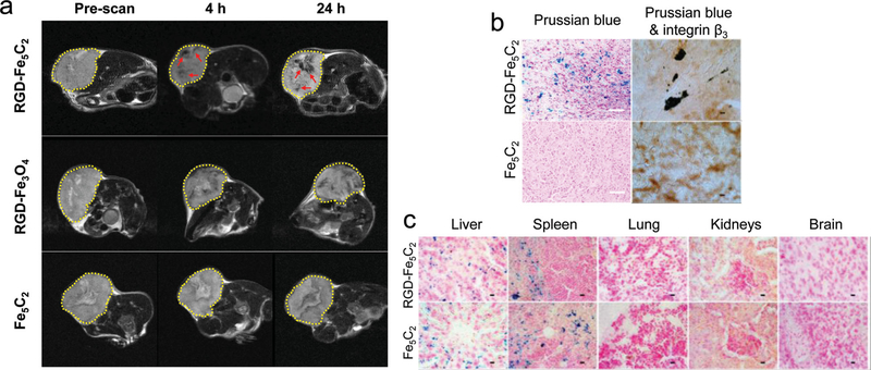
a) MR imaging results. U87MG tumor bearing mice were intravenously injected with RGD-Fe5C2, RGD-Fe3O4, or Fe5C2 nanoparticles (10 mg Fe/kg). Scans were performed before, and 4 and 24 h after the administration. In both the RGD-Fe5C2 and RGD-Fe3O4 groups, black areas (in the RGD-Fe5C2 group highlighted by red arrows) were found distributed in the tumors (circled by yellow dotted lines). RGD-Fe5C2 induced more significant signal drop than RGD-Fe3O4 at both time points. b) Left column: Prussian blue staining with tumor tissues. Positive staining was found in the RGD-Fe5C2 group but not in the Fe5C2 group. Scale bars, 50 μm. Right column: Prussian blue and integrin β3 double staining with the tumor tissues. Good overlap was observed in the RGD-Fe5C2 group, indicating that the tumor accumulation was mainly caused by RGD-integrin interactions. Scale bars, 10 μm. c) Prussian blue staining with tissue samples from the liver, spleen, lung, kidneys, and brain. Particles were accumulated in the liver and spleen but not in the other major organs. Scale bars, 10 μm.

