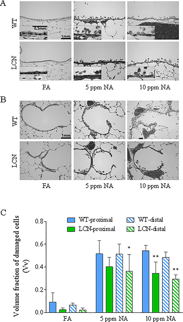
Fig. 3. High-resolution histologic analysis of the extent of airway epithelial damage induced by NA inhalation exposure in WT and LCN mice.
Two-month old wild-type (WT) and liver-Cpr-null (LCN) male mice were exposed to 5 or 10 ppm of naphthalene (NA), or to filtered air (FA), for 4 h. Mice were euthanized 20 h after termination of exposure, and lungs were processed for histopathologic analysis as described in Materials and Methods. (A, B) Typical signs of NA-induced epithelial Club cell damage, swelling, vacuolization, and exfoliation in the proximal bronchiole (A) and distal/terminal bronchiolar airways (B), as compared to the normal structures in FA-exposed control mice. Inset in A: enlarged views of swollen cells with darker nuclei staining, which signals cell death, and intracellular vacuoles. Bar = 1 mm. (C), Results of blinded quantitative analysis of the extent of cellular injury in the various groups. The volume fractions (Vv) of damaged epithelial cells are shown as means ± S.D. (n =5 for NA-exposed groups, n=4–7 for FA-exposed groups). Vv was calculated as described in Materials and Methods. All NA-treated samples show significant increase over corresponding FA samples (all p<0.0001; not labeled); *, **, p<0.05 and p<0.01, respectively, significant difference by genotype (LCN vs WT) (two-way ANOVA, followed by Bonferroni’s multiple comparisons test).

