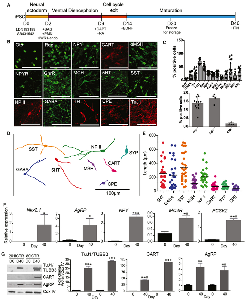
Figure 2. Characterization of Neuropeptidergic Hypothalamic Neurons.

(A) Schematic of differentiation protocol for iHTNs.
(B) Immunocytochemical staining of iHTNs showing various neuronal, hypothalamic, and neuropeptidergic markers.
(C) Quantification of the various cell types contained in iHTN cultures (n = 8 lines).
(D) Representative cell traces of iHTNs expressing relevant markers show distinctive morphologies.
(E) Quantification of primary neurite length (n = 8 lines).
(F) qRT-PCR of hypothalamic and arcuate nucleus-specific genes showing significantly increased expression ofthe genes at day 40 of differentiation compared to day 0 (n = 3 lines).
(G) Representative immunoblots and quantified histograms showing increases in neuron numbers (TUBB3) and arcuate nucleus markers (CART and AgRP) in day 40 cultures compared to day 0 (n = 3 lines). *p < 0.05, **p < 0.01, ***p < 0.001. All statistical analysis was performed using unpaired t test.
Error bars represent SEM.
