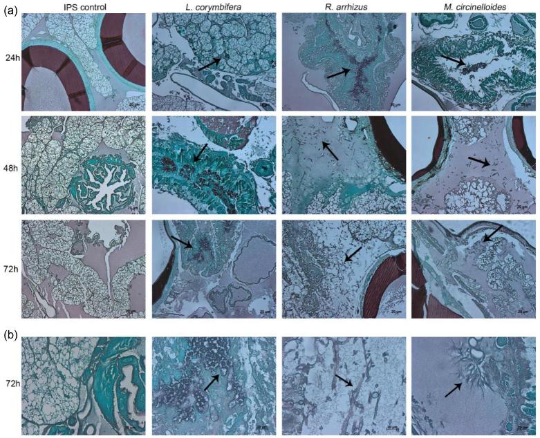
Figure 3.
Histopathology of Galleria mellonella infected with L. corymbifera AS30, R. arrhizus 44-12 and M. circinelloides AS84. (a) Larvae were infected with IPS (left panel) or 1 × 10⁶ spores of the respective strain. Larvae were fixed in formaldehyde at 24, 48 and 72 h and histological samples stained with Grocott. (b) Enlarged view of tissue sections 72 h post infection. Arrows indicate fungal elements.

