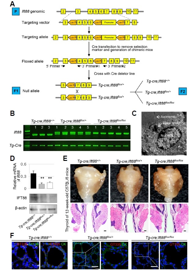
Fig. 2. Thyroid follicular cell-specific Ift88 -deficient mice exhibit ciliary loss in thyroid follicles.

(A) Generation of thyroid follicular cell-specific Ift88 knockout mice. (B) Analysis of the Ift88 genotype by PCR from mouse tail DNA. (C) The presence of primary cilia in thyroid follicular cells of Ift88 wild-type control mice was confirmed by ultrastructural analysis. (D) The expression levels of Ift88 mRNA and protein were significantly decreased in Tg-Cre;Ift88 flox/+ and Tg-Cre;Ift88 flox/flox compared with those in the Tg-Cre;Ift88 +/+ thyroid. β-actin was used as the loading control. **P < 0.01. (E) Gross morphology and H&E-stained coronal sections of the thyroid in Ift88 -deficient mice and their wild-type littermates. The thyroid glands from Ift88-deficient mice were composed of irregularly shaped follicles of variable size. The control thyroid consisted of homogeneous round follicles. Scale bar: 200 μm. (F) IF analysis of primary cilia using GT335 (red) for axoneme with basal body, Arl13B (green) for axoneme. In the Tg-Cre;Ift88 +/+ thyroid, primary cilia are present in follicular cells. In Tg-Cre;Ift88 flox/flox and Tg-Cre;Ift88 flox/+ thyroid follicles, primary cilia are almost absent. Scale bar: 10 μm.
