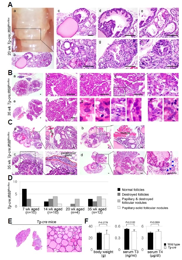
Fig. 6. Papillary-solid proliferative follicular lesions in the murine thyroid with defective Ift88 -mediated ciliary loss resemble thyroid carcinoma.

(A) At 20 weeks of age, the gross morphology of the Tg-Cre;Ift88 flox/flox thyroid showed a bubble wrap-like appearance (a). Histologically, it showed cystically dilated follicles containing dense colloid (b, c). Dilated follicles with papillary epithelial hyperplasia show scant colloid or clumped Tg globules (dh). (B) At 35 weeks of age, the Tg-Cre;Ift88 flox/flox thyroid showed proliferative nodules with papillary-solid growth patterns (a–f). The follicular epithelial cells had nuclear pseudoinclusions (g) and prominent macronucleoli (h). (C) At 35 weeks of age, the proliferative follicular nodules expanded into the perithyroidal skeletal muscle or parathyroid gland (a–c). Vascular invasion of tumor emboli was also observed (d). Scale bar: 100 μm. (D) The phenotypes of the thyroid in Tg-Cre;Ift88 flox/flox mice were summarized by age group. (E) No significant differences in morphology and function were observed between the Tg-Cre transgenic and wild-type control thyroid glands. (F) Body weight was 28.5 ± 1.0 g in wild-type mice and 26.37 ± 6.89 g in Tg-Cre transgenic mice. Serum T3 concentration was 0.53 ± 0.02 ng/ml in wild-type mice and 0.47 ± 0.04 ng/ml in Tg-Cre transgenic mice. Serum T4 concentration was 4.29 ± 0.19 μg/dl in wild-type mice and 4.66 ± 0.41 μg/dl in Tg-Cre transgenic mice.
