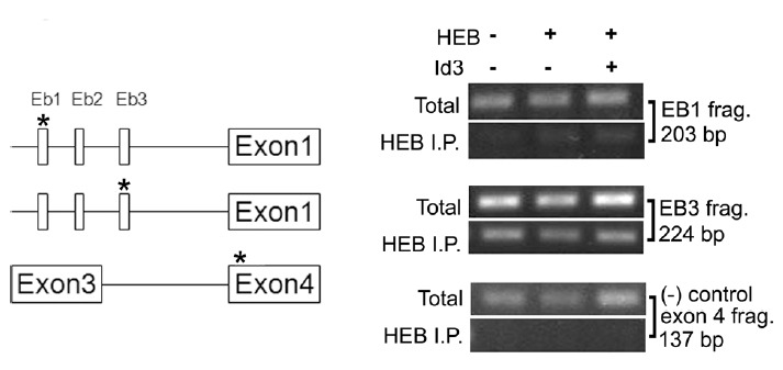
Fig. 6. Chromatin immunoprecipitation analysis of HEB binding at E-boxes of the mouse Lpar1 promoter in neocortical neuroblast cells.

Results of ChIP assay using an antibody for HEB in TR cells are shown. The location of oligonucleotide fragments used for each E-box is displayed on the map to the left. Chromatin immunoprecipitation assays were performed as described in Materials and Methods. Cells were transfected for 24 h with HEB or the Id3 expression vector (+) or without (−) before the experiments. In the ChIP assay, the Lpar1 promoter was analyzed by quantitative PCR with primer pairs. As controls, chromatin samples were analyzed before immunoprecipitation (total) to show the equal amount of starting material. * Denotes the location of the oligonucleotide fragments used for the ChIP assay in the genome map.
