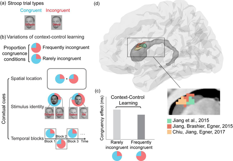
Figure 1. Context-control learning protocols and the caudate nucleus.

(a) In a face-name Stroop task, participants are asked to identify the face and ignore the overlaid written name. Congruent trials are trials where the name matches the face whereas incongruent trials are trials where the name does not match the face, thus creating conflict. The difference in response time between incongruent and congruent trials is an index of the efficiency of cognitive control.
(b) In a typical context-control learning study, there are two proportion congruence conditions – one “frequently incongruent” condition where there are more incongruent trials (e.g., 75% incongruent trials) and another “rarely incongruent” condition where there are fewer incongruent trials (e.g., 25% incongruent trials). These proportion congruence conditions can be linked to various stimulus contexts, including spatial location, stimulus identity (item-based context-control) and time frames defining temporal contexts (e.g., blocks of trials).
(c) Behavioral signature of context-control learning. The bar graph illustrates the canonical behavioral finding where there is a reduced mean congruency effect (response time for incongruent minus congruent trials) for contexts that signal more incongruent trials (frequently incongruent) than for those that signal fewer incongruent trials (rarely incongruent). This type of finding suggests that participants learn to associate a more focused attentional state with the contextual cue signaling the frequently incongruent condition.
(d) Three recent fMRI studies observed blood-oxygen level dependent (BOLD) signals in the caudate nucleus of the basal ganglia that specifically tracked long-term and short-term temporal, and item-based context-control learning processes (Chiu & Egner, 2017; Jiang et al., 2015a; Jiang et al., 2015b).These findings, based on distinct experimental manipulations and analytic approaches, converge on a zone of the caudate that is thought to form part of the “associative” cortico-striatal loop, and which receives inputs from the dlPFC (Redgrave et al., 2010).
