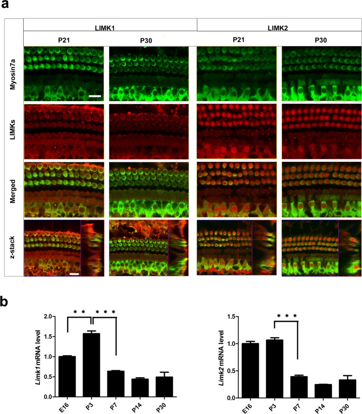
Figure 1.
Expression of LIMKs in WT mouse cochlea. (a) Immunofluorescence staining showed that LIMK1 and LIMK2 were expressed in the cochlear epithelium in the P21 and P30 mice. Myosin7a was used as a marker for HCs. Images were taken from the basal turn of the sensory epithelium. There was no difference in the immunolabeling of LIMK1 and LIMK2 from the apical to basal turns. Scale bar = 10 µm. (b) RT-qPCR results show the changes in expression of Limks in the mouse cochlea from embryonic development to adult. β-actin was used as the internal control. Data are presented as mean ± SD. (p < 0.01, n = 5).

