Figure 3.
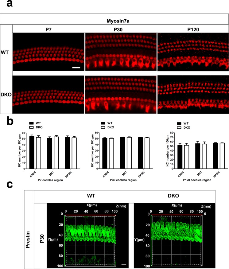
The auditory HCs are morphologically normal in the DKO mice. (a) Auditory HCs of P7, P30, and P120 mice were stained with antibodies against myosin7a and imaged using a confocal microscope. Scale bar = 10 µm. Images were taken from the basal turn of the cochlea. There was no difference in the staining from the apical to basal turns. (b) The HCs were counted and compared with age-matched WT mice (p > 0.05, n = 4). Data are presented as mean ± SD. (c) Auditory OHCs of P30 mice were stained with antibodies against prestin and imaged using a confocal microscope. Images were taken from the basal turn of the cochlea. Scale bar = 10 µm.

