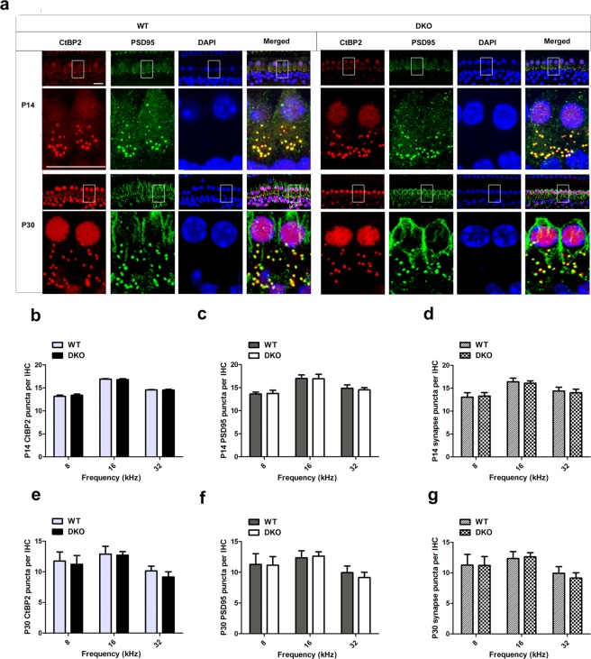
Figure 5.
The ribbon synapses were normal in DKO mice. (a) Ribbon synapses of P14 and P30 DKO and WT mice were stained with the ribbon synapse-specific markers CtBP2 and PSD95 and imaged under a confocal microscope. Images were taken from the basal turn of the cochlea, and there was no difference from the apical to basal turns (b,c,e,f). The total numbers of synapses from the 8-kHz to 32-kHz region were counted and compared between WT and DKO mice (d,g). The numbers of functional synapses in DKO mice were compared with WT mice. No significant differences were seen for any measurements (p > 0.05, n = 4). Scale bar = 10 µm. Data are presented as mean ± SD.

