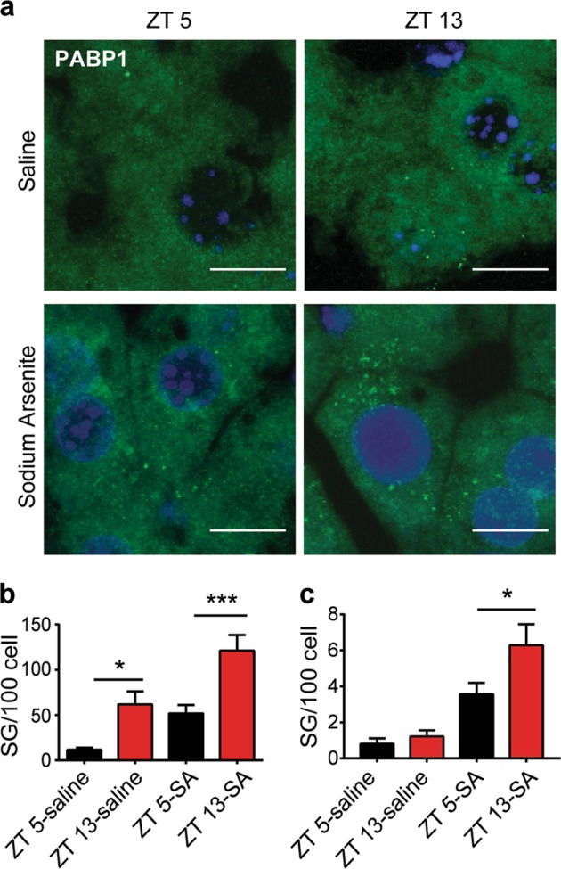
Fig. 1. Stress granules in mouse liver at zeitgeber time (ZT) 5 and ZT 13.

a The liver tissues from the wild-type (WT) mice harvested at ZT 5 and ZT 13 were labeled with anti-PABP1 (stress granule marker) antibody and revealed by immunofluorescence. Mice were intraperitoneally injected with saline or sodium arsenite (SA, 10 mg/kg) for 1 h before killing. Representative micrographs showing the presence of PABP1-positive stress granules (Scale bar = 10 μm). b, c Quantification of all visible PABP1 puncta (b) or large size puncta (>0.5 μm, c) respectively (n = 3 mice per group with 950–1000 cells examined per mouse. mean ± S.E.M.; two-way analysis of variance (ANOVA) with Tukey’s multiple comparison *P ≤ 0.05, ***P ≤ 0.001)
