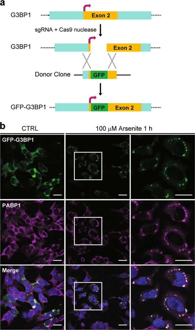
Fig. 2. Generation of GFP-G3BP1 knock-in (KI) cell line.

a Schematic representation of the strategy of tagging green fluorescent protein (GFP) at the N terminus of human G3BP1 using CRISPR/Cas9 (clustered regularly interspaced short palindromic repeats/CRISPR-associated protein 9). b Immunofluorescence confocal microscopy showing the co-localization of endogenous SG marker PABP1 with knock-in GFP-G3BP1. Stress granules were induced with 100 μM sodium arsenite for 1 h. The square areas in the middle panels were enlarged and shown in the right. Scale bar = 20 μm
