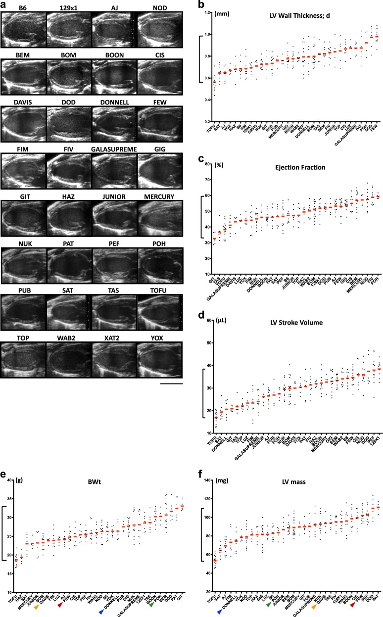
Fig. 1.
Comparative morphological and functional analysis of selected CC RI strains (12-week-old male mice). a Representative echographic images of the left ventricles in longitudinal axis. b–f Plots showing distribution of individual values among RI strains for b LV wall thickness in diastole, c ejection fraction, d LV stroke volume, e body weight (BWt), and f LV mass (calculations based on echographic measurements). Colored arrows point at the strains with most prominent differences between BWt and LV weight. Scale bar, 5 mm a

