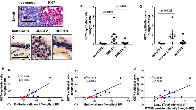
Figure 3.
Index of epithelial cell proliferation positively correlate with PTCH1 protein in moderate COPD patients. (A,B) FFPE-human tonsil sections were stained with Ki67 as an index of cellular proliferation. Representative image of human lung tissues from (C) non-COPD, (D) COPD GOLD STAGE 2, and (E) COLD GOLD STAGE 3 stained with Ki67 antibody were shown. Airway epithelial-specific Ki67+ cells was normalized to the length of basement membrane (µm) in (F) non-COPD, COPD GOLD STAGE 2 and GOLD STAGE 3,4, and (G) with COPD stratified by smoking status (current vs ex-smokers). Values were expressed as mean ± SEM in panels F and G. The Kruskal–Wallis test with Dunnett’s multiple comparisons test was used in panel F and G. Correlations between airway epithelial-specific Ki67+ cells with (H) total epithelial cell count, (I) epithelial thickness and (J) total epithelial-specific PTCH1 protein expression (data log-transformed) were shown. Linear regression analyses were used in panels H–J. Red dot = non-COPD, blue dot = COPD GOLD STAGE 2.

