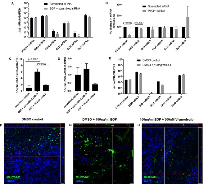
Figure 5.
EGF-induced MUC5AC expression is mediated through PTCH1-SMO axis in vitro. (A) PTCH1, SMO, SHH, GLI1, GLI2 and GLI3 mRNA expressions were assessed in NCI-H292 cells treated with EGF only. (B) Percent change in PTCH1, SMO, SHH, GLI1, GLI2 and GLI3 mRNA expressions were assessed in NCI-H292 cells treated with scrambled or PTCH1 siRNA for 48 h. (C) MUC5AC and D) MUC5B mRNA expressions were assessed in cells pre-treated with scrambled or PTCH1 siRNA followed by EGF stimulation. Values were expressed as mean ± SEM (N = 3 independent experiments). A two-tailed unpaired parametric t-test was used for each gene in panels A,B. One-way analysis of variance with Bonferroni’s multiple comparisons test was used in panels C,D. (E) PTCH1, SMO, SHH, GLI1, GLI2 and GLI3 mRNA expressions were assessed in primary bronchial epithelial cells differentiated in ALI (N = 1 replicate). Immunofluorescence images showing goblet cell expression (MUC5AC) with nuclei counterstain (DAPI) in (F) DMSO vehicle, (G) DMSO + 100 ng/ml EGF, H) and 100 ng/ml EGF + 300 nM vismodegib (SMO inhibitor) (N = 1 replicate). Cross-sections through both axes of the membrane are shown by the red (x-axis) and white (y-axis) lines.

