Figure 6.
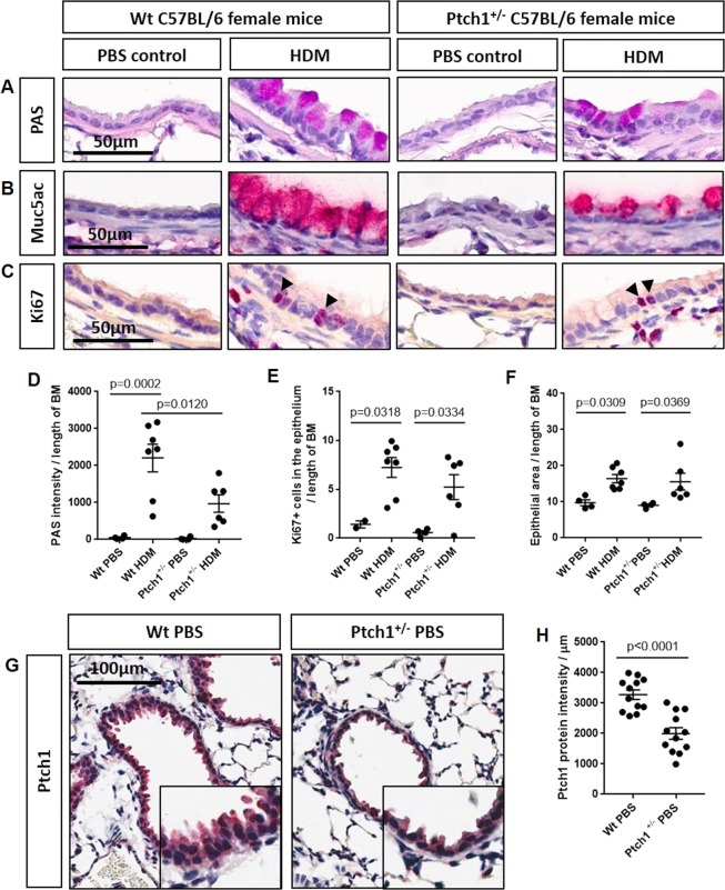
Ptch1+/− mice were partially protected from HDM-induced mucous expression compared to HDM-exposed Wt mice. (A) Periodic acid Schiff (PAS)-, B) MUC5AC- and (C) Ki67-stained sections of paraffin-embedded mouse lung tissues from Wt and Ptch1+/− mice after PBS or HDM exposures were shown. (D) Airway-specific PAS staining was normalized to length of basement membrane (BM). (E) Ki67+ epithelial cells and (G) airway epithelial thickness were normalized to length of BM. Scale bar = 50 µm. Values were expressed as mean ± SEM. One-way analysis of variance with Bonferroni’s multiple comparisons test was used in panels D–F. (G–H) Representative images and quantification of epithelial-specific Ptch1 protein expression in all visible distal airways in a representative Wt PBS- and Ptch1+/− PBS-treated mouse.

