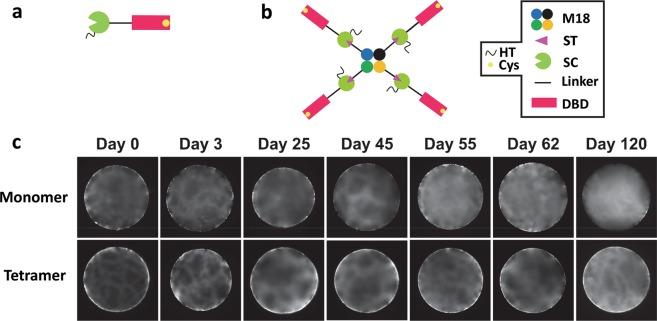
Figure 5.
Binding of the fluorescently labelled monomeric His-SC-L-DBD(Cys) and tetrameric M18-L-ST·His-SC-L-DBD(Cys) to Sephadex G-100 beads. (a,b) Domain organization of His-SC-L-DBD(Cys) and M18-L-ST·His-SC-L-DBD(Cys). HT, His-tag; Cys, cysteine; M18, SAVSBPM18; ST, SnoopTag; SC, SnoopCatcher; DBD, Dextran-binding domain. (c) Spatial and temporal distribution of fluorescently labelled monomeric His-SC-L-DBD(Cys) and tetrameric M18-L-ST·His-SC-L-DBD(Cys) to Sephadex G-100 beads. Labelled protein binding to Sephadex was analyzed up to 120 days after mixing the labelled proteins with the beads. Surface distribution of the fluorescently labelled molecules is the major focus of these pictures.

