Figure 5.
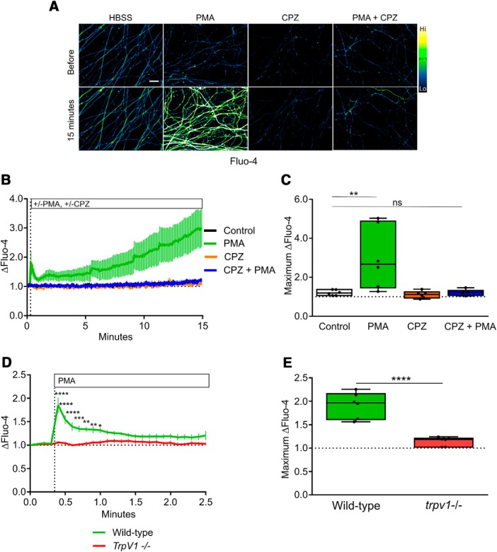
TRPV1 mediates PKC-dependent axonal Ca2+ flux. A, Representative images of DRG axons loaded with Fluo-4 and live-imaged during stimulation with PKC activator PMA in the presence or absence of TRPV1 inhibitor CPZ (10 μM). TRPV1 inhibition abolished the axonal Ca2+ response to PKC activation. B, Time course of the Fluo-4 responses to Ca2+ influx during the 15-min recording period (n = 6, mean and SEM are indicated). C, Maximum responses during the recording period were analyzed using two-factor ANOVA and Tukey’s post hoc comparison; indicated are median, min/max, and 25/75%. D, The axonal Ca2+ response to PKC activation was absent in axons of TrpV1-knock-out DRG (n = 6, mean and SEM are indicated and data were analyzed by two-factor ANOVA and Sidak’s post hoc comparison). E, The maximum Fluo-4 responses to axonal Ca2+ were analyzed by an unpaired, two-tailed t test and indicated are median, min/max, and 25/75%; *p < 0.05, **p < 0.01, ***p < 0.001, ****p < 0.0001.

