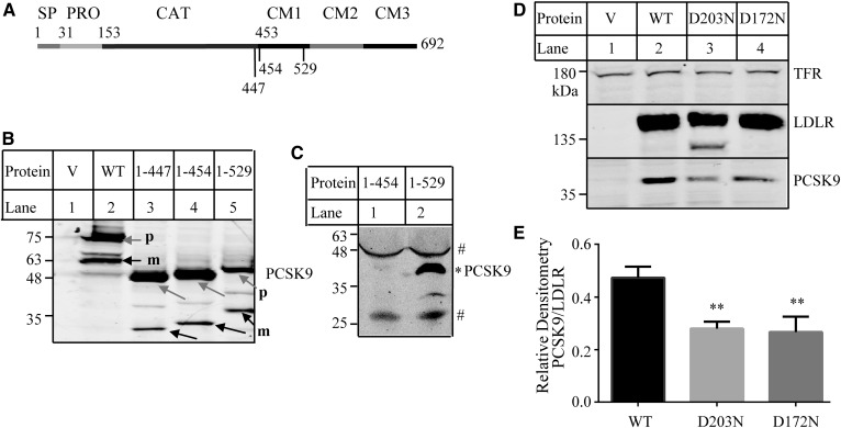
Fig. 3.
Binding of mutant PCSK9 to the WT and mutant LDLR. A: A schematic of PCSK9. Signal peptide (SP; amino acid residues 1–30); prodomain (PRO; amino acid residues 31–152); catalytical domain (CAT; amino acid residues 153–452); and C-terminal domain (amino acid residues 453–692) with three modules: module 1 (CM1; amino acid residues 457–527), module 2 (CM2; amino acid residues 534–601), and module 3 (CM3; amino acid residues 608–692) (45). B, C: Expression of the WT and mutant PCSK9 in HEK293 cells (B) and culture medium (C). HEK293 cells transiently expressing the WT or mutant PCSK9 as indicated were collected for the preparation of whole-cell lysate. Culture medium was collected from one 150 mm dish of HEK293 cells transiently transfected with mutant PCSK91–454 or PCSK91–529 and then concentrated using a 3 kDa cutoff centrifugal concentrator (Millipore). The same amount of total proteins of whole-cell lysate (B) or the same amount of concentrated medium (C) were subjected to immunoblotting using a monoclonal anti-PCSK9 Ab 13D3 that recognizes the catalytical domain of PCSK9. m, cleaved mature form of PCSK9 (black arrows); p, the precursor form of PCSK9 (gray arrows). # nonspecific bands; * mature form of PCSK91-529. D: Binding of PCSK91–529 to the WT and mutant LDLR. The experiments were performed as described in the Fig. 1 legend. Briefly, HEK293 cells transiently expressing the WT or mutant LDLR were incubated with the same amount of concentrated medium containing PCSK91–529 on ice at pH 7.4 for 4 h. LDLR and PCSK9 were detected by HL-1 and 13D3, respectively. Transferrin receptor (TFR) was detected by its specific monoclonal Ab. V: Cells were transfected with the empty vector, pCDNA3.1. E: Quantified PCSK9 binding data. The relative densitometry of PCSK9 was the ratio of the densitometry of PCSK9 to that of the mature form of LDLR. Values are mean ± SD of three experiments. ** P < 0.01.

