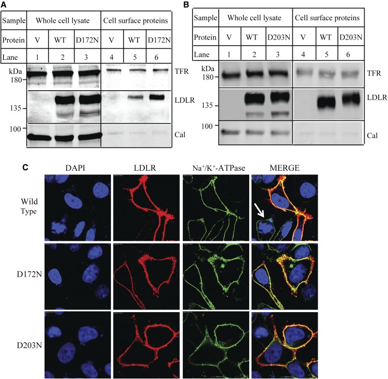
Fig. 6.
Cellular localization of the WT and mutant LDLR. A, B: Biotinylation of cell surface proteins. HEK293 cells transiently expressing the WT or mutant LDLR were incubated with Sulfo-(LC)-NHS-biotin. The whole-cell lysate was then prepared and subjected to Neutravidin agarose to pull down biotinylated cell surface proteins. LDLR was detected by HL-1. Calnexin (Cal) and transferrin receptor (TFR) were detected by their specific Abs. V: Cells were transfected with the empty vector, pCDNA3.1. C: Confocal microscopy. HEK293 cells transiently expressing the WT or mutant LDLR were fixed, permeabilized, and then incubated with a monoclonal anti-LDLR Ab and a polyclonal anti-Na+-K+-ATPase Ab. Ab binding was visualized with Alexa 568-conjugated goat anti-mouse IgG (red) and Alexa 488-conjugated goat anti-rabbit IgG (green). Nuclei were visualized with DAPI and shown as blue. An x-y optical section of the cells illustrates the distribution of the WT and mutant proteins between plasma and intracellular membranes (magnification: 100×).

