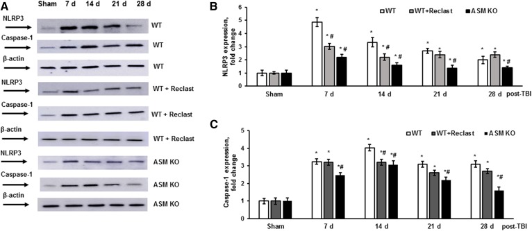
Fig. 7.
TBI-triggered NLRP3 inflammasome activation is attenuated in the ASM-deficient brain. Brain tissue samples were prepared from the injured brain of WT, ASM KO, and WT mice treated with Reclast (2 mg/kg, ip). Sham-injured animal brain (sham) was used as a control. A: Equal amount of sample (30 μg) was loaded into the lane. The expression of protein markers of the NLRP3 inflammasome were characterized by Western blotting. To confirm equal loading of samples, the membranes were stripped and probed with anti-β-actin antibody. Representative data are from six independent experiments. Quantification of the normalized NLRP3 protein expression (B) and the normalized protein expression of caspase-1 (C) using ImageJ software. Data are means ± SE. * P < 0.05 (n = 6 compared with sham); #P < 0.05 (n = 6, WT versus WT+ Reclast or ASM KO).

