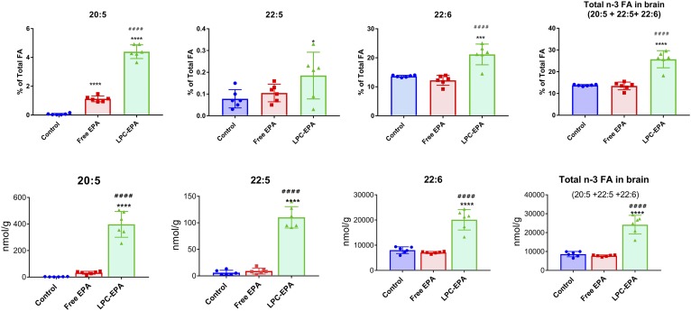
Fig. 2.
Omega 3 FA incorporation into brain lipids. A: Percent of total FAs. Total FA analysis of brain lipids was carried out by GC/MS. Only the values for the three long-chain omega 3 FAs are shown. The composition of all the FAs are shown in supplemental Table S2. The values shown are mean ± SD (n = 6 per group). Statistical significance was determined by one-way ANOVA with post hoc Tukey test. B: Nanomoles per gram of tissue. The concentration of the three long-chain omega 3 FAs in total brain lipids is expressed as nanomoles per gram. The statistical symbols are the same as in Fig. 1.

