Fig. 7.
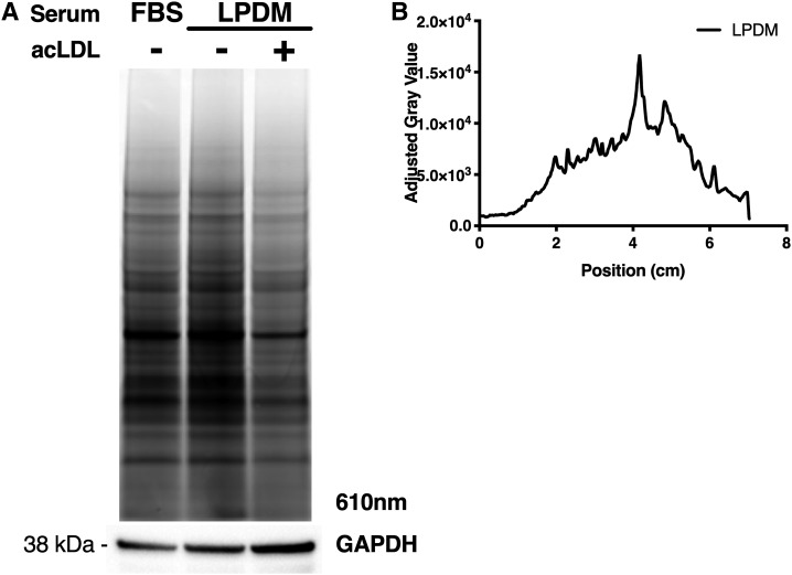
Fluorescent gel profiling reveals specific labeling of proteins. Following treatment of U2OS-SRA cells with 10 μM probe in complete media, cells were incubated with complete media (FBS), LPDM, or LPDM containing 100 μg/ml acLDL. After 18 h, cells were cross-linked, lysed, and clicked to rhodamine X azide before separation by SDS-PAGE. A: Representative gel imaged at 610 nm, with parallel immunoblot for GAPDH. B: Gel profile quantified using ImageJ software. The graph quantifies the difference in the gray value between LPDM with and without acLDL as a measure of competition due to excess cholesterol.

