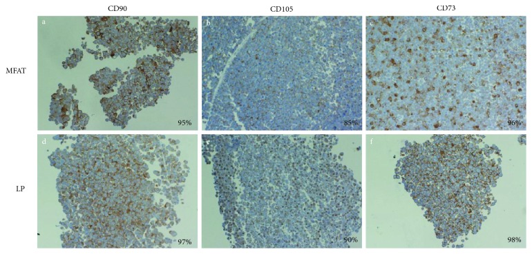
Figure 2.
Expression of CD90, CD105, and CD73 markers in CD31 cells derived from MFAT and LP specimens. CD31 cells isolated from MFAT and LP specimens were cultured for 14 days. At the end of incubation, the cells were recovered, cytoincluded in Matrigel, and analyzed by immuocytochemistry. The figures (10x magnification) shows MFAT (a, b, and c) and LP (d, e, and f) staining for CD90, CD105, and CD73, respectively. The % of positive cells is reported at the lower right corner of each picture.

