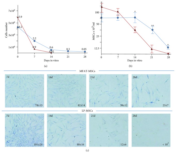
Figure 3.
Total cell number and MSC content in LP and MFAT specimens. An identical volume (3 ml) of MFAT (blue line) and LP (red line) specimens was digested with collagenase upon cultivation in DMEM plain medium for 0, 7, 14, 21, and 28 days to evaluate the total cells (a) and MSC content (b). On day 0, the total cells/ml as well the MSC content were higher in LP than in MFAT (total cells LP = 6.3 ± 4.4 × 10⁵ vs. MFAT = 3.7 ± 1.8 × 10⁵; MSCs LP 14.9 ± 6.3 × 10³ vs. MFAT 10.1 ± 5.8 × 10³). After 14 days of culture, both total cells and MSCs were significantly reduced in LP, whereas in MFAT remained stable. (c) Pictures (20x magnification) of MSCs isolated and seeded in T25 flask, from LP and MFAT specimens. At the lower right corner of the pictures, the cell number/field is reported and represent the average ± SD of 5 different fields. Eight different donors were analyzed.

