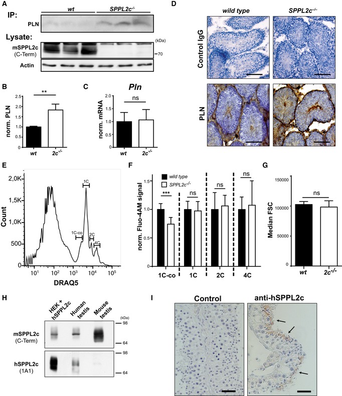
Figure 5. SPPL2c regulates Ca2+ homeostasis in spermatids by cleaving phospholamban.

-
AAccumulation of endogenous PLN in the testis of SPPL2c −/− mice. PLN was immunoprecipitated (IP) from equal protein amounts of total lysates of wild‐type or SPPL2c −/− testis with the mouse monoclonal antibody. Western blot analysis of the IPs was performed with the rabbit monoclonal PLN antibody. SPPL2c and actin were detected from total lysates directly.
-
BQuantification of normalised PLN/Actin ratios from n = 3 biological replicates per genotype. Mean ± SD, unpaired Student's t‐test; **P ≤ 0.01.
-
CExpression of PLN in murine testis from wild‐type (wt) SPPL2c −/− (2c −/−) was quantified by qPCR. n = 6. Unpaired Student's t‐test. ns = not significant.
-
DImmunohistochemical detection of PLN in paraformaldehyde (PFA)‐fixed testis from wild‐type and SPPL2c −/− mice. Cryosections were stained using a rabbit monoclonal PLN antibody or normal rabbit IgG as negative control and diaminobenzidine as peroxidase substrate. Scale bars, 100 μm.
-
ETestis suspensions were stained with 2.5 μM DRAQ5 to determine cellular DNA amount. 1C‐co = elongated spermatids, 1C = other haploid cells including round spermatids, 2C = spermatogonia, secondary spermatocytes, Sertoli cells, other somatic cells, 4C = primary spermatocytes, G2 spermatogonia.
-
FIntracellular Ca2+ concentrations were analysed in testis suspensions from either wild‐type or SPPL2c‐deficient mice using the Ca2+‐sensitive probe Fluo4‐AM. Individual germ cell populations were identified as shown in (E). Bars indicate Median Fluo4‐AM fluorescence ± SD of 8 individual samples from three mice per genotype normalised to those of wild‐type samples. Unpaired Student's t‐test. ***P ≤ 0.001, ns = not significant.
-
GMedian forward scatter (FSC) was controlled in the 1C‐co population from the data sets shown in (F). Median FSC ± SD of 8 individual samples from three mice per genotype. Unpaired Student's t‐test. ns = not significant.
-
H, IExpression of SPPL2c in human testis. (H) Western blot analysis of lysates from HEK293 cells expressing human (h) SPPL2c, human and murine testis using antisera against a C‐terminal epitope of murine SPPL2c or human SPPL2c as indicated. (I) Immunohistochemical detection of SPPL2c with the monoclonal 1A1 antibody in paraffin sections from Bouin's‐fixed human testis. In addition, sections were incubated with control immunoglobulins. Arrows indicate elongated spermatids. Scale bars, 30 μm.
Source data are available online for this figure.
