-
A
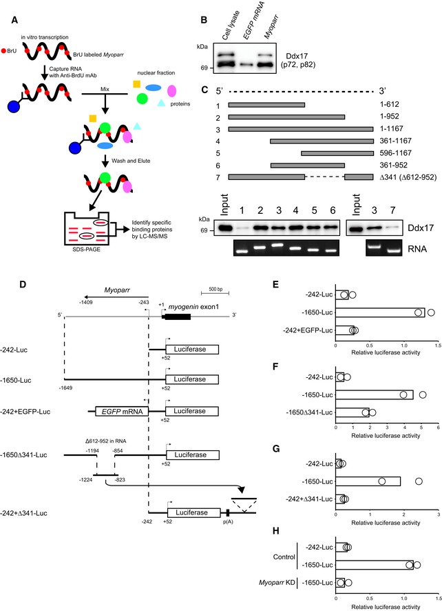
Scheme of the identification of Myoparr‐interacting proteins using RiboTrap and differential proteomics analysis.
-
B
Following immunoprecipitation, the interaction between Myoparr and endogenous Ddx17 was confirmed by immunoblotting using a Ddx17‐specific antibody. Two different Ddx17 isoforms (p72 and p82) were observed.
-
C
Determination of the Ddx17‐binding region of Myoparr by RNA pull‐down analyses. Schematic diagram of full‐length or truncated Myoparr used for RNA pull‐down (top). In vitro‐transcribed/in vitro‐translated Ddx17 protein was pulled down by indicated Myoparr and then detected by Western blot using a Ddx17 antibody (bottom).
-
D
Schematic diagram of the constructs used for luciferase assays. The details of all constructs are described in
Materials and Methods. p(A) indicates poly(A) site.
-
E–G
Relative luciferase activities of indicated constructs in differentiating C2C12 cells.
-
H
Relative luciferase activities of indicated constructs in differentiating C2C12 cells transfected with control or Myoparr shRNA (Myoparr KD).
Data information: In (E‐H), bars indicate the average of two independent experiments, and open circles represent the values of each experiment.

