
Figure EV3. ER and Golgi morphology in HEK293 cells with ectopic expression of SPPL2c.
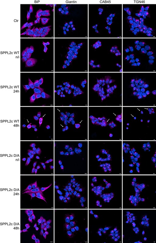
Cells were seeded and cultured for 72 h and expression of catalytically active (WT) or non‐active (D/A) SPPL2c was induced by addition of doxycycline for either 24 or 48 h as indicated. Non‐transfected cells (Ctr) or non‐induced SPPL2c cells (n/i) served as controls. The ER was stained with the anti‐BiP antibody, the cis/medial‐Golgi with the anti‐Giantin, the trans‐Golgi network with the anti‐TGN46 antibody, and all Golgi subcompartments with the CAB45‐specific antibody. Note that only upon expression of catalytically active SPPL2c, the morphologies of ER and cis‐Golgi change compared to controls. The most representative changes are indicated with white arrows at SPPL2c WT 48 h. This figure contains a smaller magnification of the same experimental setup as in Fig 5D. Scale bar 5 μm.