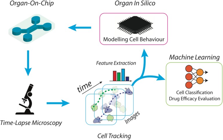
FIGURE 1.
Scheme of a high throughput platform for the advanced study and reproduction of the tumor microenvironment. The microfluidic device is manufactured ad hoc, according to the biological experiment requirements. Then, the desired cell subsets are loaded into the chip together with tumor cells, to an extent to propose a simplified version of the tumor microenvironment. Time-lapse microscopy is used to acquire the high-resolution frames of the whole video sequence. Microscopy setting is functionalized by the scale of the objects of interest and the duration of the time-lapse. Cells are then automatically localized and tracked across each frame of the video sequence and trajectories are characterized in terms of individual and aggregated kinematics and morphological descriptors. At this point, specifically developed machine learning algorithms are then applied to recognize patterns for biological reasoning. For example, cell tracking datasets are then clustered into separated groups reflecting distinct cell behaviors. The same kinematics and morphological descriptors can be used as input for in silico models aimed at simulating on-chip experiments.

