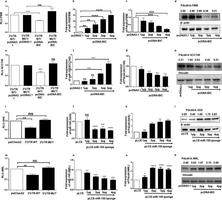
FIG 2.
Targeting of 3′ UTR of Pdcd4 by miR-155 and the consequent changes in the expression of Pdcd4 in FBM, SAS, and AWL cells. (a and e) A dual-luciferase reporter assay was performed in FBM and SCC745 cells by cotransfecting them with the Pdcd4WT or MUT 3 UTR (100 ng) and pcDNA3.1 or pcDNA-BIC. These graphs represent normalized values (relative light units [RLU]) of Renilla/firefly luciferase activities (n = 3). (b and f) Expression of miR-155 normalized to that of U6 in FBM and SCC745 cells transfected with different amounts (1 μg to 4 μg) of pcDNA-BIC with pcDNA3.1 as a control (n = 3). (c and g) Expression of Pdcd4 mRNA in FBM and SCC745 cells transfected with different amounts (1 μg to 4 μg) of pcDNA-BIC with pcDNA3.1 as a control and normalized to that of β-actin mRNA (n = 3). (d and h) Western blots for Pdcd4 and β-actin in FBM cells and for Pdcd4 and vinculin in SCC745 cells transfected with different amounts (1 μg to 4 μg) of pcDNA-BIC, with pcDNA3.1 as a control. (i and m) Dual-luciferase reporter assay performed in SAS and AWL cells transfected with 100 ng of psiCheck-2, psiCheck-Pdcd4-WT 3′ UTR or psiCheck-Pdcd4-MUT 3′ UTR (n = 3). (j and n) Expression of miR-155 in SAS and AWL cells transfected with pLCE–miR-155 sponge plasmid (1 to 4 μg), with 4 μg of pLCE as a control (n = 3). (k and o) Expression of Pdcd4 mRNA in SAS and AWL cells transfected with pLCE–miR-155 sponge plasmid (1 to 4 μg) with 4 μg of pLCE as a control, normalized to that of β-actin (n = 3). (l and p) Western blots for Pdcd4 in SAS and AWL cells when transfected with 1 μg to 4 μg of pLCE–miR-155 sponge plasmid, with 4 μg of pLCE as a control; β-actin and vinculin were taken as internal controls, respectively. Values are expressed as the means ± SD (*, P < 0.05; **, P < 0.01; ***, P < 0.001; ****, P < 0.0001; ns, not significant).

