
Figure 1.
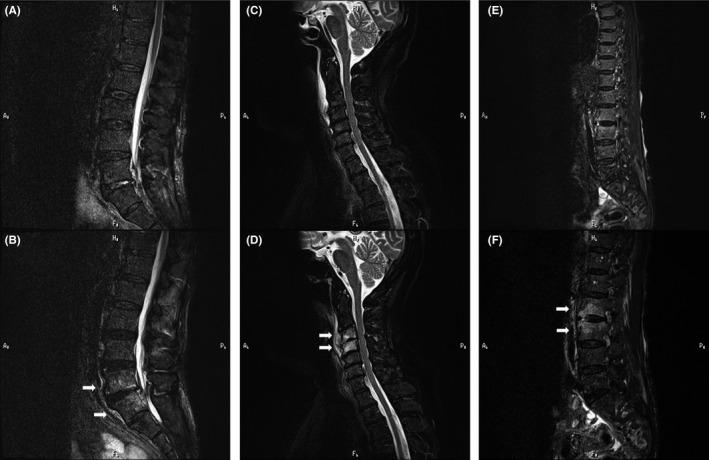
Initial magnetic resonance (short time inversion recovery) images obtained <2 wk after the onset of symptoms showed no obvious abnormalities: A, case 1; C, case 2; and E, case 3. Follow‐up magnetic resonance images performed 2‐4 wk after the onset of symptoms detected high‐intensity lesions in the vertebral bodies. Arrows indicate the lesion: B, case 1; D, case 2; and F, case 3