
Fig. 1. Post-transplant hepatic oxidative stress was milder in mice receiving Ppara-null (−/−) livers than in those receiving wild-type (+/+) livers.
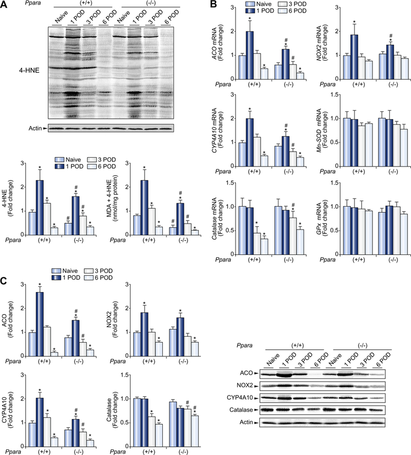
(A) Immunoblot analysis of 4-HNE and quantification of MDA + 4-HNE levels in the liver. Whole liver lysates (100 μg of protein) obtained from one mouse in each group were loaded onto the gels. Actin was used as the loading control. Band intensity was quantified densitometrically, normalized to that of actin, and subsequently normalized to that of naïve Ppara+/+ mice. Measurement of hepatic MDA + 4-HNE is described in Materials and methods. Results are expressed as mean ±SD(n = 4in each group). POD, post-operative day; *p <0.05 vs. naïve mice of the same genotype; #p <0.05 vs. Ppara+/+ mice at the same time point. (B) Hepatic mRNA levels of ROS-generating and ROS-eliminating enzymes. The mRNA levels were normalized to those of GAPDH mRNA, and subsequently normalized to those of naïve Ppara+/+ mice. Results are expressed as mean ± SD (n = 4 in each group). Abbreviations are the same as panel (A). (C) Immunoblot analysis of selected ROS-related enzymes. The same samples used in panel (A) (whole liver lysate, 50 μg of protein) were loaded onto the gels. Band intensity was quantified densitometrically, normalized to that of actin, and subsequently normalized to that of naïve Ppara+/+ mice. Results are expressed as mean ±SD (n = 4 in each group). *p <0.05 vs. naïve mice of the same genotype; #p <0.05 vs. Ppara+/+ mice at the same time point.