
Fig. 2. Post-transplant hepatic mitochondrial FAO enzymes and PDK4 were down-regulated in mice receiving wild-type (+/+) or Ppara-null (−/−) livers.
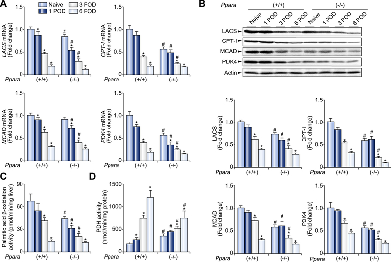
(A) Hepatic mRNA levels of mitochondrial FAO enzymes and PDK4. The mRNA levels were normalized to those of GAPDH mRNA, and subsequently normalized to those of naïve Ppara+/+ mice. Results are expressed as mean ±SD (n = 4 in each group). POD, post-operative day; *p <0.05 vs. naïve mice of the same genotype; #p <0.05 vs. Ppara+/+ mice at the same time point. (B) Immunoblot analysis of mitochondrial FAO enzymes and PDK4. The same samples used in Fig. 1A (whole liver lysate, 50 μg of protein) were loaded onto the gels. Band intensity was quantified densitometrically, normalized to that of actin, and subsequently normalized to that of naïve Ppara+/+ mice. Results are expressed as mean ±SD(n = 4in each group). Abbreviations are the same as panel (A). (C and D) Activities of mitochondrial β-oxidation and PDH. Results are expressed as mean ±SD (n = 4 in each group). Abbreviations are the same as panel (A).