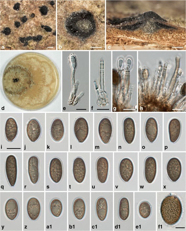
Fig. 3.
Juglanconis pterocaryae. a Conidiomata in surface view. b, c Transverse (b) and vertical (c) sections of conidiomata, showing central column. d Culture (CMD, 25 d, 16 °C). e–h Conidiophores (annellides; in e, g with young conidia). i–e1 Vital conidia with gelatinous sheath. f1 Squashed conidium showing the densely verruculose inner conidial wall. All in water (a–c, i–m, f1 WU 39981, neotype; d WU 39983; e, f, n–x WU 39982; g, h WU 39985b; y–d1 WU 39986b; e1 WU 39987a). Scale bars a 500 μm; b, c 200 μm; e–e1 10 μm; f1 5 μm

