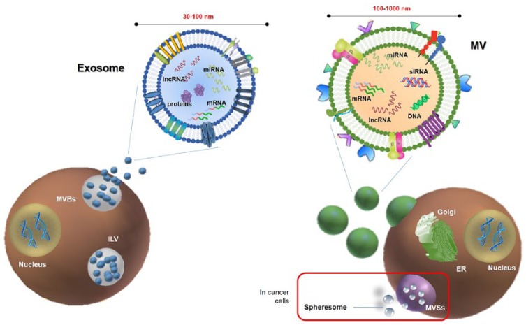
Figure 2.
Schematic representation of extracellular vesicle (EV) release. Left panel: Exosomes (30–100 nm in size) are released in extracellular space from multivesicular bodies (MVBs) through exocytosis. MVBs contain various intraluminal vesicles (ILVs) which are generated by the inward budding of the endosome membrane. Exosome cargo may include different kind of RNAs, such as miRNA, lncRNAs, mRNAs, otherwise quickly degraded if free. Right panel: Microvesicles (100–1000 nm in size) originate through a finely regulated budding/blebbing of the plasmatic membrane involving the Golgi apparatus. According to the classical secretory pathway, vesicles with their protein cargo, are sorted and packed in the Golgi apparatus, and then transported to the plasma membrane. In cancer, it has been proposed that there is an additional mechanism of EV release. Specifically, cancer cells may produce multivesicular spheres (MVSs), which contain many spheresomes.

