Fig. 1.
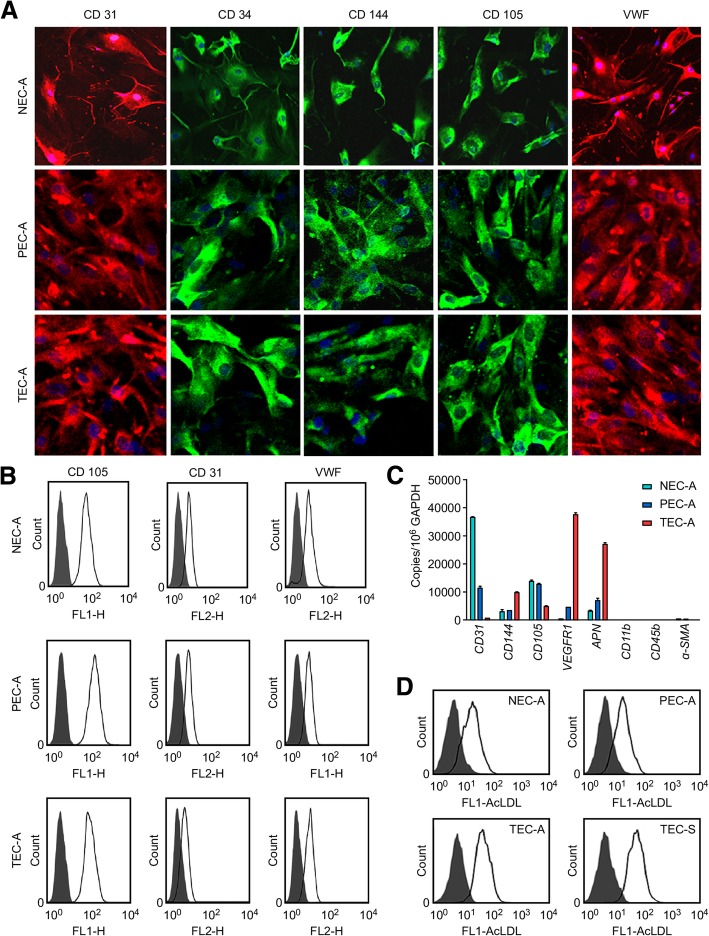
Assaying the expression of endothelial markers and AcLDL uptake in normal tissue-, paratumor- and tumor-derived ECs. a Positive immunoreactivity with antibodies specific for CD31 (red), CD34 (green), CD144 (green), CD105 (green), and VWF (red) using NEC-A (normal), PEC-A (paratumor), and TEC-A (tumor) tissues from six patients with lung ADC. Nuclei were stained with DAPI (blue). b Expression of the EC markers CD31 and VWF and the microvascular EC marker CD105 in NEC-A, PEC-A, and TEC-A tissues was evaluated by flow cytometry. c The mRNA expression levels of CD105, CD31, CD144, VEGFR1, APN, CD11b, CD45b, and α-SMA in NEC-A, PEC-A, and TEC-A tissues. The data were normalized to GAPDH expression and shown as the fold change compared to NECs. d Alexa Fluor® 488 AcLDL uptake assays. Uptake efficiency in NEC-A, PEC-A, TEC-A, and TEC-S (from tumor tissues of six lung SCC patients) was detected by flow cytometry

