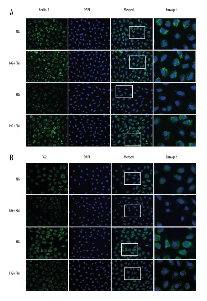
Figure 3.
Effects of PM on the expression of Beclin-1 (A) and P62 (B) were determined by immunofluorescence test. HK-2 cells were cultured in NG and then stimulated by HG for 48 hours. Beclin-1 and P62 (green) was defined by staining cells with anti-Beclin-1 and anti-p62 antibody. DAPI staining (blue) was used to determine the nucleus position. Values are expressed as means ± standard deviation. NG – normal glucose, 5.5 mM glucose; HG – high glucose, 30 mM glucose; NG+PM – PM (1 mM) plus 5.5 mM glucose; HG+PM – PM (1 mM) plus 30 mM glucose. (# P<0.05, ## P<0.01 compared with NG. * P<0.05, ** P<0.01 compared with the HG group). PM – pyridoxamine; HK-2 – human proximal tubular epithelial cells; DAPI – 4,6-diamino-2-phenyl indole.

