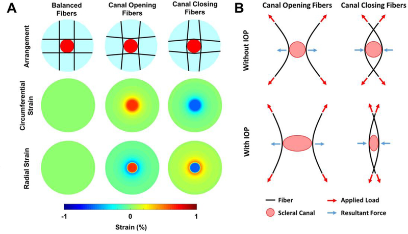
Fig. 8.

Small changes in the orientation of the fibers determined whether the canal opened or closed due to IOP-induced hoop stress. A) Strain maps for the different fiber arrangements. When the fibers were oriented convexly to the canal (Canal Opening Fibers) the strains in the lamina were positive. When the fibers were oriented concavely around the canal (Canal Closing Fibers) the strains in the lamina were negative. B) Diagram depicting the mechanism of action. For fibers oriented concavely around the canal, the applied load results in an outward tensile force at the canal boundary as the fiber straightens. For fibers oriented convexly around the canal, the applied load results in an inward compressive force at the canal boundary as the fibers straighten. Note only two fibers for each case are shown here for simplicity.
